Written by: Mike McLean
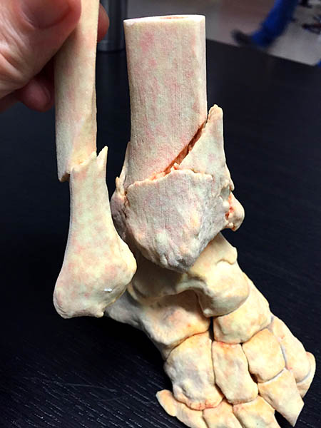
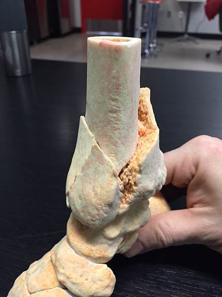
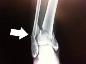
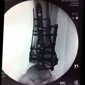
In December of 2014, I was coaching my sons hockey team when I lost an edge and fell hard, crashing into the boards at full speed. The resulting fracture in my lower leg resembled the trauma that may be found in a head-on collision or a fall from 30 feet. The Tibia and Fibula had both broken, with the base of the Tibia taking the worst of it, shattering into 14 pieces. The world class medical team at Spectrum Health’s Butterworth campus quickly developed a surgical strategy which included 2 operations, 4 plates and over 20 screws. While the resulting surgeries were declared successful, I was left wondering, how were the surgeons able to piece together the myriad fragments and what strategy was used to locate and secure the necessary hardware? The answers were explained to me through the help of a Color Jet Printed recreation of my original CT scan.
A lot has been written about the benefits of 3D printing; how it can democratize design, speed product development cycles or replace conventional machining techniques. Medical applications of 3D printing in general seem to get a fair amount of press; we’ve seen stories ranging from prostheses for pets to tracheal splints in pediatric surgery. One of the oft overlooked benefits of 3D printing is the enhanced communication that a physical object can bring to the table.
“This is exactly how the site looked,” said Dr. Terrence Endres, orthopaedist with Orthopaedic Associates of Michigan, as he examined the 3D print. Orienting the Tibia model to align with my outstretched leg, he explained how each surgical scar corresponded to a major bone fragment, that was secured to the intact bone via a stainless steel plate. Once secured, each larger piece in turn acted as the anchor upon which to base the reduction of the smaller pieces. Dr. Endres pulled out his smartphone and snapped a few quick pictures of the model, clearly impressed by the accuracy of the representation.
While Color Jet Printing technology is greatly suited for patient education, other additive manufacturing technologies lend themselves to more advanced applications. Cardiovascular surgical planning has been enhanced through Multi-Jet Modeling, in which tissue density and durometer are accurately modeled in a lifelike representation of the heart. Such models are helping to reduce time in the OR and lessen liability for the staff. Surgical implants and tools have been created through Direct Metal Printing, Stereolithography and Selective Laser Sintering, with each technology featuring multiple bio-compatible materials.
As patient specific medicine continues to trend upward, expect to see additive technologies playing a key role in elective surgeries and educational tools; eventually becoming a necessary extension of medical imaging, where the technology will continue to have a growing impact on positive outcomes in the surgical suite and ultimately the bottom line.
If you ever find yourself looking at the business end of a scalpel, you can drift off knowing that somewhere along the chain of medical technology, 3D printing played a part. Until then, keep your head up and your stick on the ice.